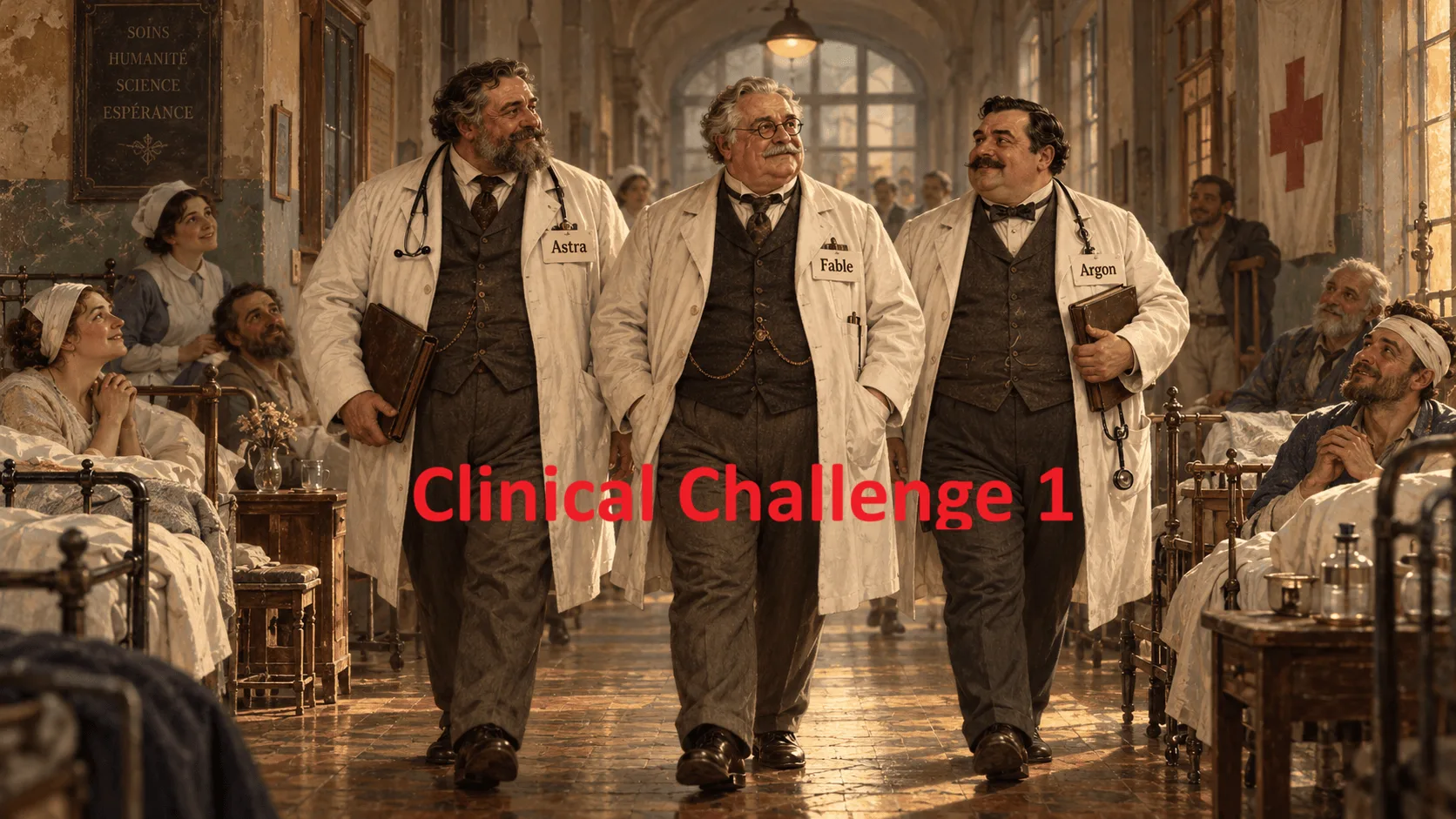
Series: The AI Giants Enter the CardiacSurgery ICU — “I Big entrano in corsia” – Clinical Challenge 1
A mean arterial pressure of 76 mmHg. Cardiac index 1.7 L/min/m². Lactate still rising. Has the patient improved?
In the first episode I set GPT-6 Astra, Claude Fable 5.1 and Gemini 4 Argon a small clinical challenge, and their preliminary scores came out almost identical. Scores like that are easy to report and much harder to interpret. So this time I want to look past them, at what the answers say about actual clinical decisions.
The first case is one in which blood pressure and perfusion move in opposite directions.
A few ground rules. The patient is fictional. Every value, and the order in which the values appear, was fixed before any model saw the case.
T0 — An apparently familiar postoperative problem
The patient is in the first hours after coronary artery bypass grafting on cardiopulmonary bypass. MAP 61 mmHg, heart rate 88/min, cardiac index 3.0 L/min/m², low systemic vascular resistance, ScvO₂ 72%, lactate 1.7 mmol/L. Bedside echocardiography shows preserved left and right ventricular systolic function and no significant effusion. Norepinephrine is running at a low dose.
Nothing here would surprise anyone who works in a cardiac ICU.
Your decision: What is the priority diagnosis, what should be done now, what additional information matters most, and what remains uncertain?
What the three models said at T0
GPT-6 Astra read the picture as post-bypass vasodilatory hypotension compatible with vasoplegia, not as low-output cardiogenic shock. Its advice: titrate norepinephrine promptly toward an individualized perfusion-pressure target, avoid unselected fluid boluses and, if more volume is being considered, assess fluid responsiveness dynamically first.
Claude Fable 5.1 came to essentially the same physiological conclusion but put the immediate urgency at low to moderate. It focused on close observation (drain output, hemoglobin, urine output, serial lactate) and on rethinking the diagnosis if vasopressor requirements went up.
Gemini 4 Argon also saw vasodilation with preserved global perfusion indices. It wanted norepinephrine adjusted within minutes, and asked for urine output, drainage and hemoglobin to judge organ perfusion and to look for occult blood loss.
My reading: T0 does not justify a diagnosis of acute right ventricular failure, and none of the three made one. All of them kept a clear line between a low MAP caused by low resistance and a true low-output state. Where they differed was in how urgent they sounded. Not in the diagnosis.
T1 — Ninety minutes later, the physiology changes
Vasopressor support has been increased, yet MAP is now 59 mmHg. Cardiac index has dropped to 1.6 L/min/m² and ScvO₂ to 50%; lactate is up to 4.0 mmol/L. Central venous pressure 18 mmHg. On repeat echocardiography the right ventricle is dilated and severely hypokinetic, with no severe left ventricular depression. The mediastinal drains show no relevant blood loss.
Stop here: Would you treat this primarily as persistent vasoplegia? Give fluid? Add an inotrope? Investigate a surgical cause? Prepare mechanical support?
What the models said at T1
Astra dropped vasoplegia as a sufficient explanation, and said so explicitly. It called this low-output shock dominated by acute right ventricular dysfunction. Its priorities were urgent cardiac-surgical involvement, tailored vasoactive and inotropic support, no empirical fluid loading, and a parallel search for reversible causes. It pointed to transesophageal echocardiography as especially useful for picking up a localized compressive process that the first examination may have missed. (Anyone who has found a localized clot pressing on the right heart after an unremarkable transthoracic study will appreciate the point.)
Fable reached the same diagnosis, acute right ventricular cardiogenic shock, but ranked myocardial ischemia, or an acute problem with the right coronary graft, high on the differential. It proposed immediate inotropic support, reducing right ventricular afterload where possible, assessing the graft territory, and thinking early about mechanical assistance. It asked for an ECG with right-sided leads, while keeping compressive and pulmonary vascular causes on the list.
Argon called it frank right ventricular low-output shock and, like Fable, flagged ischemia or a sudden rise in pulmonary afterload. Its recommendations: emergency inotropic support, no further blind fluid administration, optimized ventilation, and immediate cardiac-surgical involvement.
What matters is that all three accepted that new data required a new working diagnosis. Whether a model put echocardiography or the ECG first is secondary. The patient had changed, and the response had to change too. The importance of challenging an initial diagnostic interpretation is also discussed in my article on multi-agent AI and diagnostic bias.
T2 — The dangerous illusion of recovery
More vasopressor, and MAP climbs to 76 mmHg. Cardiac index, however, is only 1.7 L/min/m², ScvO₂ 49%, lactate 4.8 mmol/L, central venous pressure 19 mmHg. The right ventricle is still severely compromised.
The arterial pressure is better. The circulation is not.
What the models said at T2
Astra warned that getting the MAP back is not the same as getting perfusion back. Its priority was to improve forward flow immediately: inotropic support, urgent evaluation for reversible causes, and early activation of a mechanical-support assessment if cardiac output did not pick up quickly. It also cautioned against simply turning up the vasoconstrictors, or giving fluid, without a physiological reason.
Fable used the most decisive escalation language of the three at this stage. It described the higher MAP as a misleading normalization produced by vasoconstriction and recommended activating the mechanical circulatory support pathway now (potentially VA-ECMO or right-sided assistance, depending on anatomy, physiology and local expertise) while proceeding straight to surgical reassessment for correctable causes.
Argon labeled the picture refractory right ventricular shock despite a seemingly satisfactory blood pressure. It favored immediate inotropic support over further escalation of pure vasoconstrictors, and wanted the team alerted urgently to the possibility of surgical or angiographic correction and of mechanical assistance.
This is where the comparison gets interesting. All three recognized persistent shock. They did not agree on how soon to move toward mechanical support.
The clinical issue is not the MAP alone
MAP matters for organ and coronary perfusion; nobody disputes that. But pressure is not cardiac output. Raise systemic vascular resistance and the measured arterial pressure goes up, while forward flow can stay just as poor. In this vignette everything else points the same way: a cardiac index that is still low, ScvO₂ falling, lactate rising, high filling pressure, severe right ventricular dysfunction. Reading 76 mmHg as clinical recovery would be a mistake. A related distinction between treatment intensity and physiological response is explored in my article on the Vasoactive-Inotropic Score.
The guidance is less tidy than one might wish. The Association for Acute CardioVascular Care of the ESC has published a clinical consensus statement on acute right ventricular failure (Arrigo et al.) that covers perfusion markers such as lactate and SCAI shock staging, venous congestion, echocardiographic assessment and cause-directed treatment, with a dedicated section on temporary mechanical circulatory support. One caveat, though: its scope is right ventricular failure secondary to an acute rise in afterload (acute cor pulmonale), so it maps only partly onto an ischemic or graft-related problem after CABG. On the surgical side, the 2024 EACTS/EACTAIC/EBCP guidelines on cardiopulmonary bypass deal with separation from bypass, difficult weaning and temporary mechanical circulatory support.
The document closest to this scenario is the 2020 EACTS/ELSO/STS/AATS expert consensus on post-cardiotomy extracorporeal life support (Lorusso et al.). On timing it is explicit: post-cardiotomy support should be initiated before end-organ injury or the onset of anaerobic metabolism, with a lactate below 4 mmol/L as the reference (Class I, Level B). Our patient was already at 4.0 mmol/L at T1 and 4.8 mmol/L at T2, which is worth keeping in mind when judging how early Fable wanted to escalate. On isolated right ventricular failure the same document is far more tentative: an oxygenated RVAD configuration or a short-term ventricular assist device “may be considered” (Class IIb, Level C). None of these documents turns a given lactate or CVP into an automatic indication for one particular device, and post-cardiotomy support decisions still rest on specialist assessment.
Relevant reading: ACVC/ESC clinical consensus on acute right ventricular failure, 2024 EACTS/EACTAIC/EBCP guidelines on cardiopulmonary bypass in adult cardiac surgery and 2020 EACTS/ELSO/STS/AATS expert consensus on post-cardiotomy ECLS.
My clinical commentary: three good answers, one real decision point
I would not call any of the three responses a gross clinical error. Each one picks up the evolving shock state, and none of them falls for the temptation to call the problem solved because the blood pressure now looks normal. In practice that temptation is real, particularly at the end of a long night.
Where I would draw a line is between recognizing that advanced support is needed and committing to a specific form of support immediately. Mechanical assistance may well become essential. But the choice of device, and the chance to correct a graft, ischemic, compressive or pulmonary vascular cause directly, depend on further information. Information that has to be gathered in minutes, not hours.
Fable’s firmer language can be read two ways. As commendable anticipation of where the patient is heading, or as more prescriptive than the available data allow. I am not entirely sure which reading is right. Astra and Argon leave a little more room to see how the patient responds to inotropes and to complete an urgent, cause-directed assessment, while still making it clear that escalation is needed.
One thing I want to state plainly: you cannot find the correct threshold by counting how many appropriate therapies a model mentions.
Our prewritten rubric gave all three models 5/5. For the essential clinical principles, that seems defensible to me. It does not mean the three would have made the same decisions at the bedside.
What would you have done?
Imagine you are the intensivist or cardiac surgeon responsible for this patient at T2. The arterial pressure now reads 76 mmHg. Lactate keeps rising, ScvO₂ keeps falling, and the severe right ventricular dysfunction has not changed.
Would you activate a mechanical support pathway immediately, or allow a closely monitored trial of targeted inotropic support while investigating a reversible cause? Which additional finding would change your decision?
Coming next, Episode 03: an extubated patient whose SpO₂ stays reassuring while ventilation quietly fails. When should a model give up on noninvasive support and protect the airway?
Editorial note: Fictional case; not a clinical management protocol. This commentary does not establish patient-level safety or model superiority.
References
- ACVC/ESC clinical consensus statement: right ventricular failure secondary to acutely increased afterload (acute cor pulmonale)
- 2024 EACTS/EACTAIC/EBCP Guidelines on cardiopulmonary bypass in adult cardiac surgery
- 2020 EACTS/ELSO/STS/AATS expert consensus on post-cardiotomy extracorporeal life support in adult patients